KEY ADVANTAGES
Simultaneously meet the needs of dermatologists and cosmetic surgeons
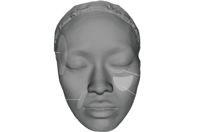
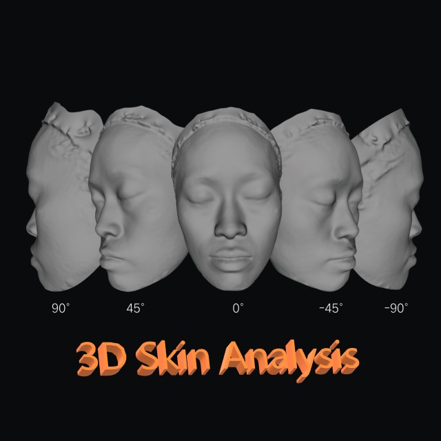
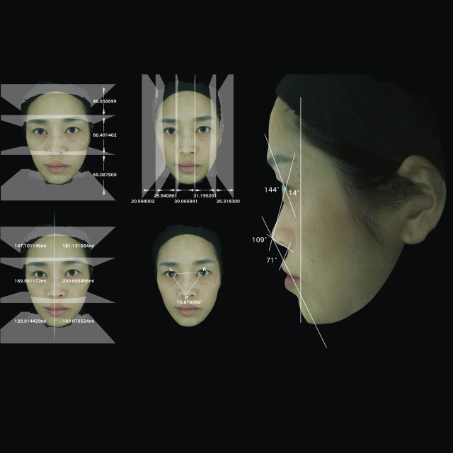
3D full-facial skin images, bid farewell to 2D aesthetic measurement, and effectively assist facial microplastic surgery consultation
Capture skin images in a true and natural state under a standard daylight color temperature of 6000K;
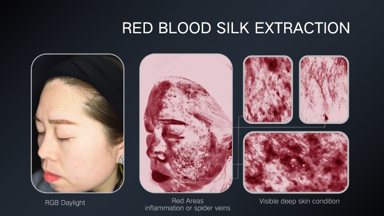
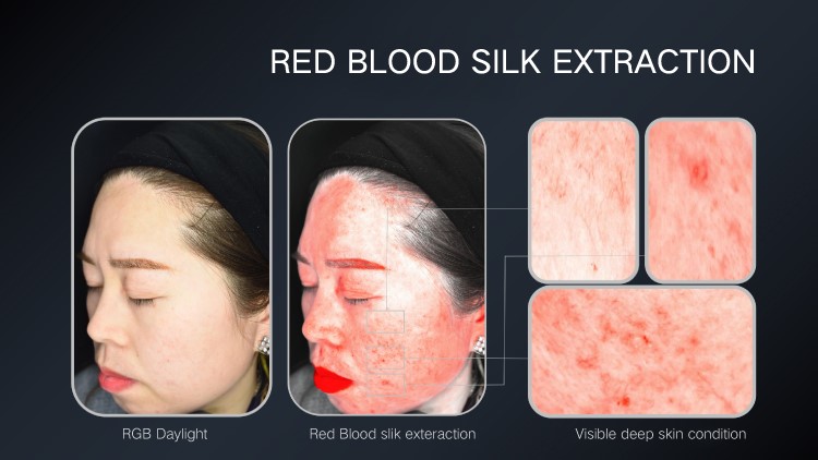
Visible spots, epidermal pigmentation, skin texture, and smoothness on the skin surface;
Wrinkles, texture, Enlarged Pores, acne marks, pigmentation, etc.
“Ultimate optical imaging technology, restoring true colors.”
Spyder CHECKR48 color correction, providing more precise adjustment and calibration for skin analysis applications, restoring the most accurate state of the skin.
AUTOMATIC ROTATION CAMERA, REACHES TO 0.1MM SCANNING ACCURACY
The automatic rotating scanning camera can shoot to obtain 0°-180° full-face images of 0.1mm accuracy. No need to adjust the posture, so as to save shooting time greatly. The easier shooting process makes the before-after comparison cases more standardized.

-
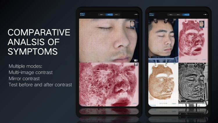
Multi-Image Comparison
Mirror Comparison: Allows for comparing symptoms on a single side of the face.
Two-Image Comparison: Enables observation of skin conditions at different time periods.
Multi-Image Comparison: Suitable for comparing skin conditions before and after long-term treatments.
3D Comparison: Shows changes in skin texture before and after treatments.
-
Symptom Annotation and Measurement
The device provides multiple tools for annotating and measuring symptoms, allowing doctors to record and save information promptly. The measurement tools are useful for comparing anti-aging and contouring treatments.
-
3D Imaging
It visualizes the skin surface in 3D from any angle, magnifying subtle skin conditions such as wrinkles, sagging, and indentations.
Software Advantages
-
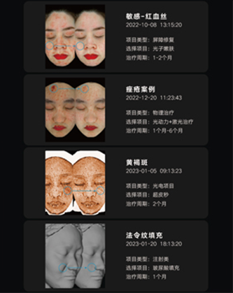
Generate Professional Case Library with One Click
The D8 skin imaging analysis device supports the rapid generation of comparative cases, while generating cases that display symptom names, care projects, lifecycle, and other relevant information. All generated cases will be automatically recorded in the system's case library.
-

Light and Shadow Diagnosis Function
By using the 360° light and shadow diagnosis function, it can more intuitively identify problems such as facial depressions and sagging.
-
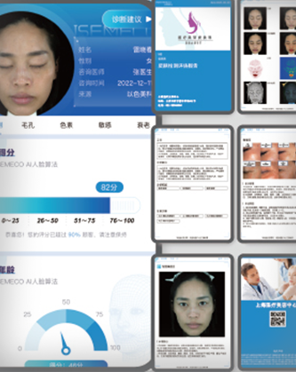
Personalized Customized Report
The D8 skin imaging analysis device supports incorporating the customer's 3D full-face image, the doctor's analysis recommendations, and recommended skincare plans into the report. This is achieved through a professionally customized report that combines images and text output.

| Name : | Model Number : |
| Skin Imaging Analyzer | D8 |
| ………………………….. | ……………………… |
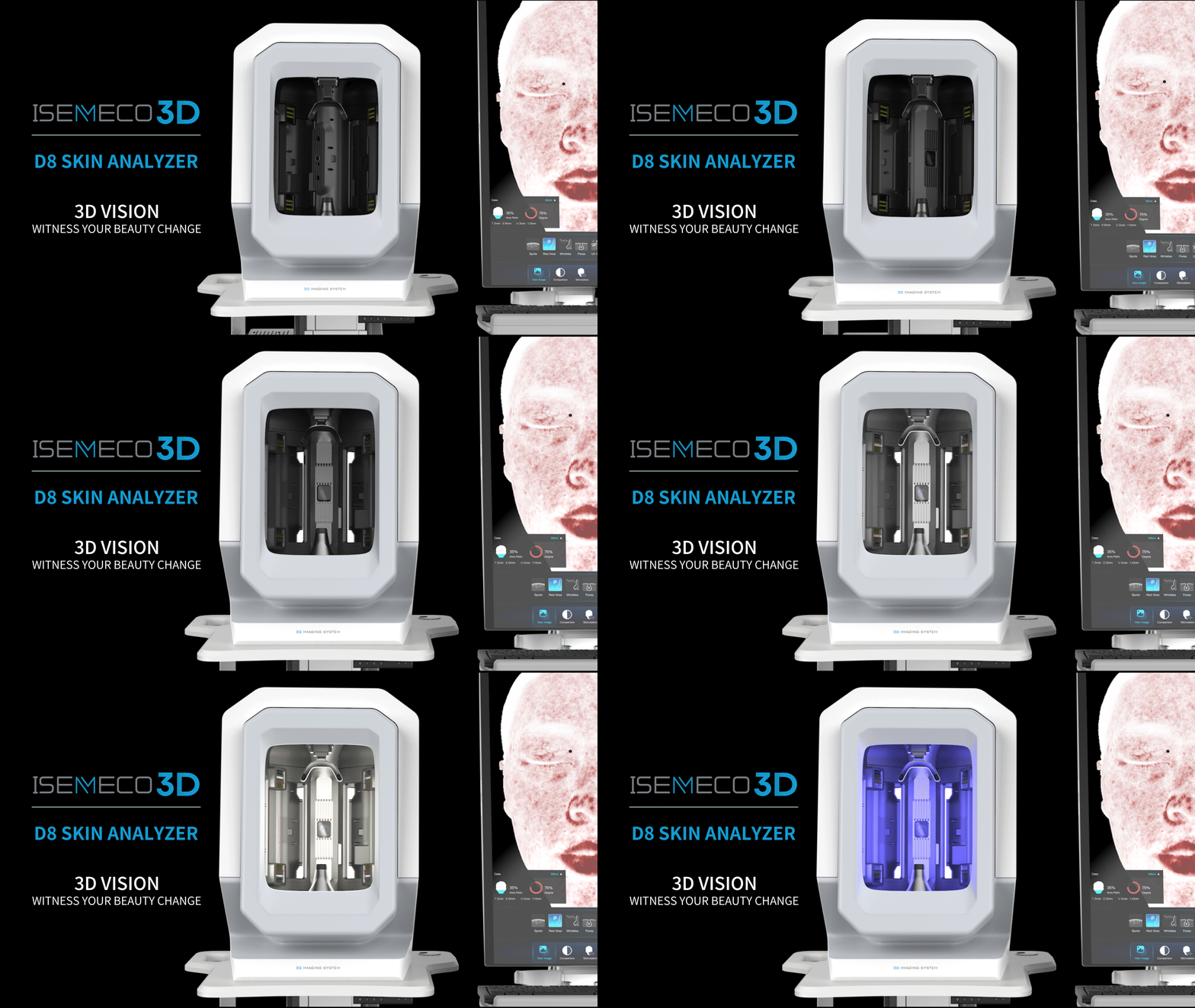
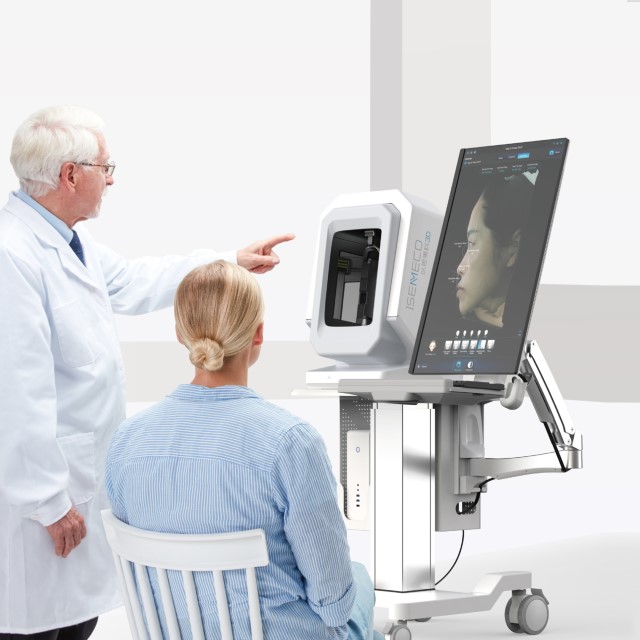
ISEMECO D8 3D Facial Skin Analyzer
|
ISEMECO Parameter |
|
| Model | ISEMECO D8 |
| Image capture | daylight , parallel polarized light, cross polarized light, and UV light. |
| Color | White |
| CMOS | 1 inch |
| Weight: | 120kg |
| Shading Method Deploy | With shading |
| Configuration | HD display+PC computer |
| Resolution | 35M Pixel |
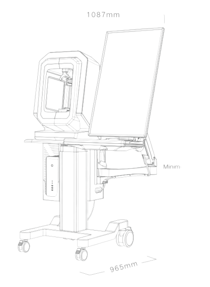
| Skin Analysis Host Size | L:1087mm W:965mm H:1470-1850mm |
| 3D modeling accuracy: | 0.2mm |
| Facial vertices: | 800,000 |
Skin Analysis Machine
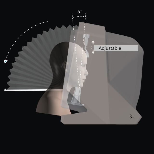
*With Concealed hood
*35million Pixel ultramicro optical lens
*UV light source imported from Japan
*Layout design of expert lightsource of chinese academy of sciences
*International standard factory production
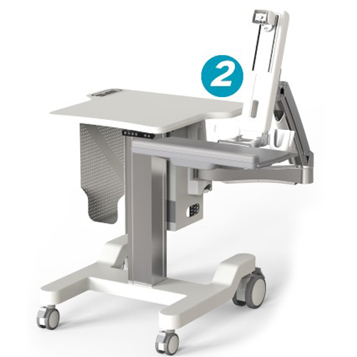
Work Table Lifting Platform
Working stand-up, relieving pressure on spine
Electric lift regulation, adjust height stable at constant speed
Height memory function for convenient use
Adjustable height for various human height
wide adjustable range of height
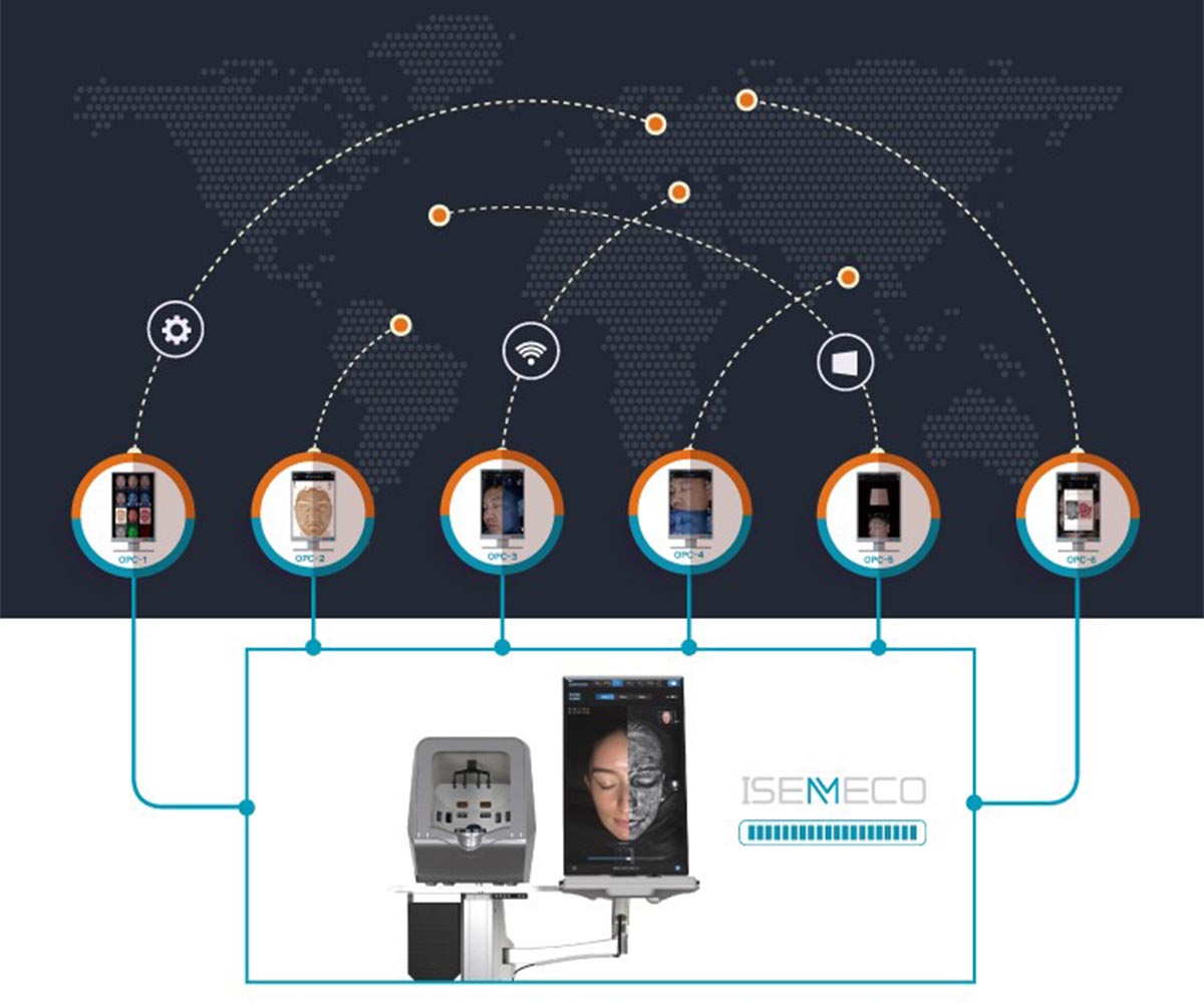
Support multi-port Access
Simultaneously meet the needs of dermatologists and cosmetic surgeons
20 Seconds, 4 spectra images of the whole face can be taken quickly.
0.1mm scanning accuracy, binocular grating structured light camera
aaS.CRM data interface


